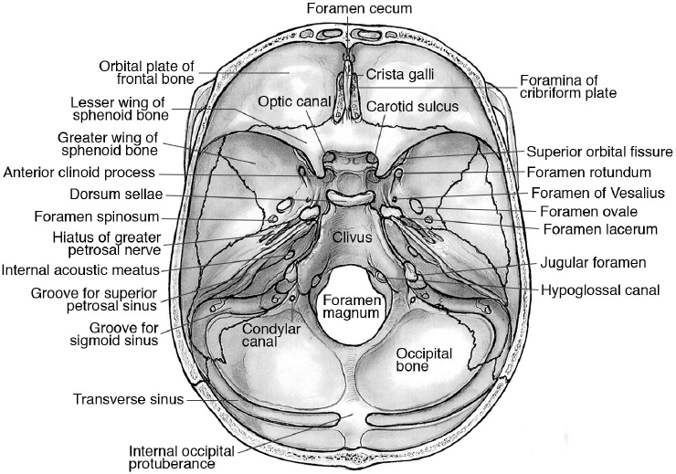
Floor View Skull Dorsum Sellae

Specialised Projections Of The Skull Radiology Key
Middle Cranial Fossa Boundaries Contents Teachmeanatomy

Landmarks For Cephalometric Analysis S Sella Center Of Sella Download Scientific Diagram
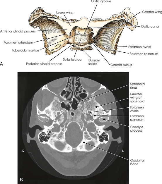
Anatomy Of The Skull Base And Related Structures Elements Of Surgical Anatomy Neupsy Key

Skull X Ray Lateral View Note Enlargement Of Pituitary Fossa Loss Download Scientific Diagram
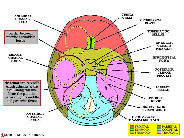
Pixelated Brain Module 1 Section 1 The Skull
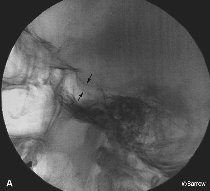
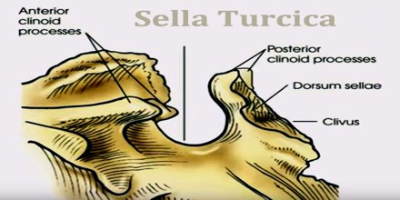
Erosion of anterior and posterior clinoids can be seen pituitary tumors e g.
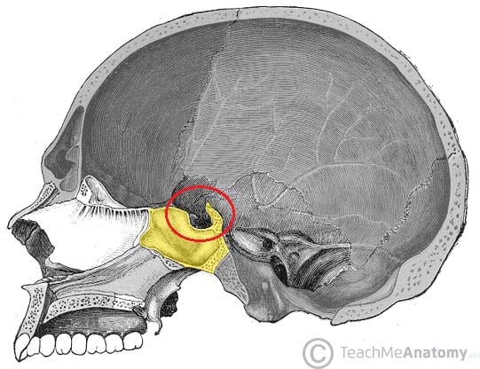
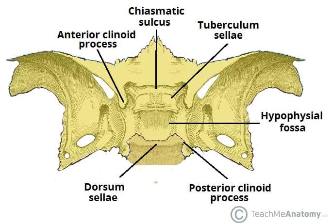
Floor view skull dorsum sellae. Together with the basilar part of the occipital bone it forms the clivus. What structure should be projected within the shadow of the foramen magnum on a well positioned ap axial towne projection of the skull dorsum sellae what is the name of the structure that houses the pituitary gland. The dorsum sellae forms the posterior wall of the sella turcica which houses the pituitary gland. The towne view is an angled ap radiograph of the skull used to evaluate for fractures of the skull and neoplastic changes.
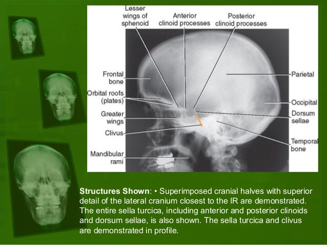
The dorsum sellae is part of the sphenoid bone in the skull. Enlargement with erosion of anterior cortex of dorsum sellae proceeds to the floor of the sella and may result in complete destruction of the dorsum. The projection is used to visualize the petrous part of the pyramids the dorsum sellae and the posterior clinoid processes which are visible in the shadow of the foramen magnum. The dorsum sellae will be projected under the foramen magnum.
Together with the basilar part of the occipital bone it forms the clivus. Skull pa axial caldwell view. The dorsum sellae is part of the sphenoid bone in the skull. It serves as a cephalometric landmark.
This is a caudally angled occipito frontal projection that demonstrates the floor of sella turcica. Under angulation over angulation shifting of the anterior or posterior clinoid within the foramen indicate what towne. Posteriorly it articulates with the basilar part of the occipital bone and jointly forms the clivus. In a properly positioned caldwell projection the ir is perpendicular to the orbitomeatal line oml and the x rays pass at an angle of 15 degrees from behind the head and exit at the nasion.

The Axial Skeleton Flashcards Quizlet
Sphenoid Bone Location Structure Function Teachmeanatomy
Skull Foramina Fissures And Contents Kenhub

Sphenoid Bone
Double Sellar Floor Radiographic Sign For A Pituitary Adenoma Barrow

Procedures 3 Skull Anatomy Flashcards Quizlet
Positioning And Radiographic Anatomy Of The Skull

Imaging Of Skull Base Lesions Sciencedirect

Anterior Cranial Fossa Nasal Cavity And Paranasal Sinuses Radiology Key
Skull Radiology Key
Sella Turcica Assignment Point
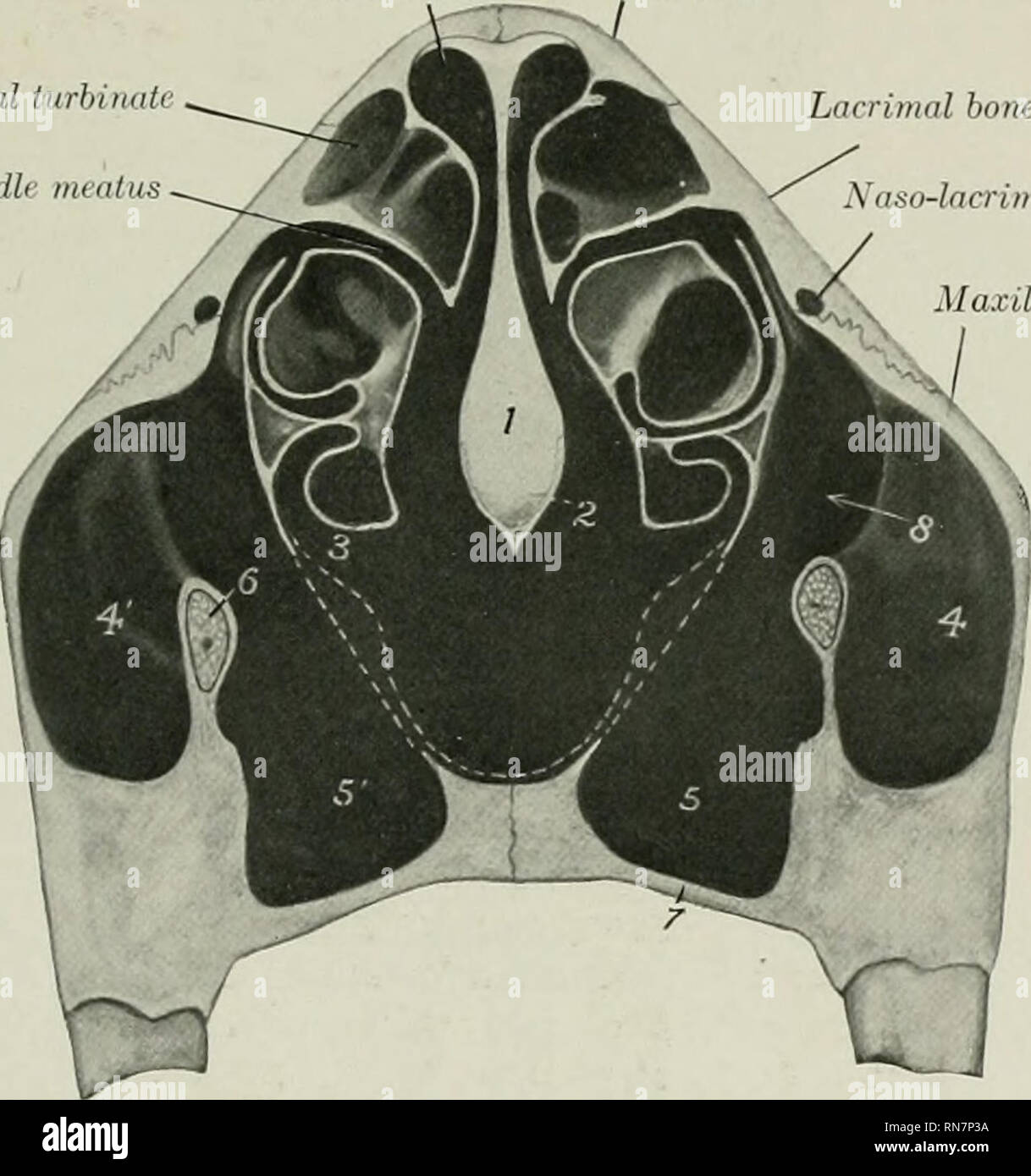
The Anatomy Of The Domestic Animals Veterinary Anatomy Skull Of The Ox As A Whole 143 Than The Rest Of The Floor The Ethmoidal Fossffi Are Smaller And The Hypophyseal Fossa

Fingerstocspine Ppt Ap Axial Skull Dorsum Sellae Projected In Foramen Magnum Entire Skull Visualized No Rotation Or Tilt Petrous Ridges Symmetric Course Hero

